How sound waves create moving pictures of your heart without using any radiation
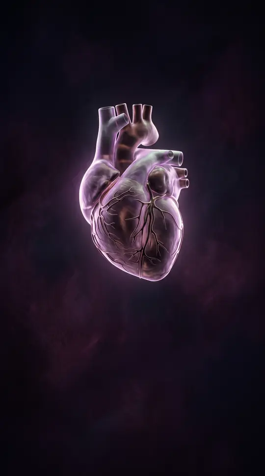
In short: Echocardiography transforms sound waves into real-time visual data, allowing doctors to observe your heart muscle and valves in motion. This painless, non-invasive scan provides critical insights into blood flow and structural health, helping specialists diagnose conditions or monitor recovery without the risks associated with traditional radiation-based imaging.
At its core, an echocardiogram functions like a medical sonar. A device called a transducer emits sound waves that bounce off heart tissue, creating echoes that computer software translates into moving images. This technology allows clinicians to assess the heart's size, shape, and pumping strength, as well as the functionality of its valves. Because it relies on ultrasound rather than ionizing radiation, it is considered a safe, standard diagnostic tool for evaluating cardiovascular health.
The most common version, the transthoracic echocardiogram (TTE), involves moving a probe over the chest after applying a conductive gel. For more complex needs, variations exist: bubble studies use saline injections to detect irregular connections between heart chambers, while stress echocardiograms monitor heart performance during physical exertion or induced elevated heart rates. In some cases, a transoesophageal echocardiogram (TOE) is used, where a transducer is passed into the oesophagus to capture higher-resolution images from a closer vantage point.
Beyond simple visualization, advanced techniques like Doppler imaging measure blood flow velocity and direction, while strain imaging tracks how muscle fibers squeeze and relax. These parameters are essential for identifying structural irregularities, such as valve leakage or narrowing, and for calculating metrics like ejection fraction. By adjusting measurements for factors like body surface area, specialists can tailor their analysis to the individual, ensuring that the diagnostic data is as accurate as possible for guiding treatment decisions.
While echocardiography is a cornerstone of modern cardiology, its utility is best realized when clinical status changes. Health societies generally advise against routine testing if a patient remains asymptomatic and the results are unlikely to alter their care plan. When used appropriately, however, it remains an indispensable tool for managing everything from heart failure and valve disease to congenital abnormalities, providing a clear, real-time window into the heart's mechanical performance.
Opens the Intellect+ feed with this card first. Swipe up for the next one.
Sources
- Summary by Intellect · source: Echocardiography
- Also: Echocardiogram
- Also: What Is an Echocardiogram?
- Also: Echocardiography
To cite this card, link to https://intellectplus.co.uk/app/health-echocardiography.
How we source, check and correct cards: Editorial standards.
General information, not professional advice. Spotted an error? Flag the card or see corrections and complaints.
Intellect Plus is a visual educational discovery platform for exploring fascinating facts and ideas. Brain Food, Not Brain Rot. What is Intellect Plus?
Related
More in Health, medicine and disease · More in Health · Latest · A–Z · All topics
