How a simple X-ray translates shadows into life-saving diagnoses
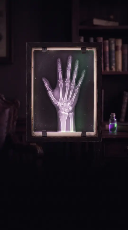
In short: When you stand before an X-ray machine, you aren't just capturing a picture; you are mapping the density of your internal world. By measuring how radiation interacts with different tissues, doctors can distinguish between the solid architecture of bone and the airy expanse of the lungs.
The mechanics of a chest radiograph (CXR) rely on differential absorption. As radiation passes through the body, dense structures like bones block most of it, appearing white on the final image. In contrast, the lungs allow more radiation to pass through, resulting in a gray appearance. Healthcare providers analyze these specific colors and shadings to identify conditions such as pneumonia, emphysema, or congestive heart failure. To ensure clarity, patients must remain perfectly still and hold their breath, as even the slight movement of breathing can blur the diagnostic data.
Technicians use specific orientations to provide a 3D understanding of the chest from 2D images. The standard posteroanterior (PA) view involves the patient facing a detector with the X-ray source behind them, typically at a distance of 6 feet (1.8m). A lateral view, where the patient stands with their side against the plate and arms raised, provides a different perspective. In contrast, anteroposterior (AP) views—where the beam enters from the front—are generally reserved for bedridden patients because they are more difficult for radiologists to interpret accurately.
While CXRs are the most common medical films, they are not always the definitive answer. They are excellent for screening, such as identifying job-related lung disease in mining, but some conditions require follow-up imaging for a definitive diagnosis. Furthermore, clinical guidelines suggest that for certain issues, like a non-displaced rib fracture, an X-ray may not be necessary as it won't alter patient management. Modern research is now pushing these boundaries, testing how high-resolution AI models can better detect small focal abnormalities that the human eye might miss.
Safety remains a primary consideration in diagnostic imaging. A chest X-ray uses a very small amount of ionizing radiation, typically resulting in a mean dose of about 0.02 mSv for a front view and 0.08 mSv for a side view in adults. To put this in perspective, the total dose for both views is roughly equivalent to the background radiation exposure one would receive over about 10 days. The risk of radiation-induced cancer from a single X-ray is estimated to be less than 1 in 1,000,000.
Opens the Intellect+ feed with this card first. Swipe up for the next one.
Sources
- Summary by Intellect · source: Chest X-ray (CXR): What You Should Know & When You Might Need One
- Also: Chest X-ray
- Also: Chest radiograph
To cite this card, link to https://intellectplus.co.uk/app/health-chest-radiograph.
How we source, check and correct cards: Editorial standards.
General information, not professional advice. Spotted an error? Flag the card or see corrections and complaints.
Intellect Plus is a visual educational discovery platform for exploring fascinating facts and ideas. Brain Food, Not Brain Rot. What is Intellect Plus?
Related
More in Morbidity · More in Health · Latest · A–Z · All topics
