The invisible space that allows your lungs to move
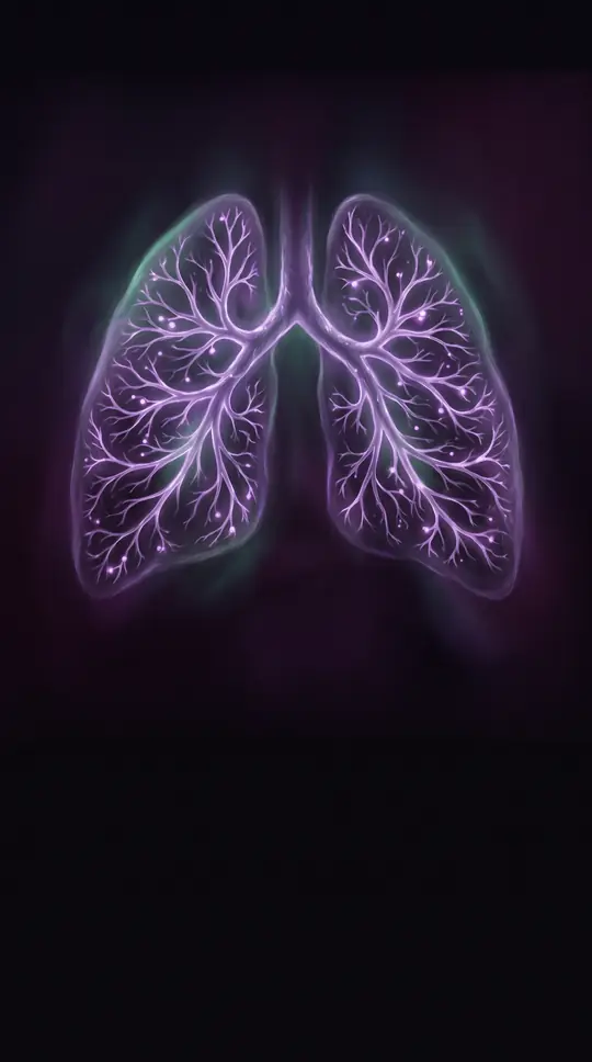
In short: Your lungs don't just expand on their own. They rely on a microscopic, fluid-filled gap between two layers of membrane. This potential space, the pleural cavity, acts as a mechanical bridge, translating the movement of your ribcage into the rhythmic expansion of your breath.
The pleural cavity is a potential space formed by two serous membranes: the visceral pleura, which covers the lungs and adjoining structures, and the parietal pleura, which lines the chest wall. These layers are separated by a tiny amount of pleural fluid—only a few milliliters in a normal human. This fluid is essential for lubrication, allowing the visceral pleura to glide against the parietal pleura without friction or sound during normal respiration. However, this system is also a mechanical transmitter; it ensures that movements of the chest wall are transmitted directly to the lungs, a process that becomes particularly critical during heavy breathing.
Anatomically, the human body maintains a strict separation between the left and right pleural cavities. There is no natural connection between them, meaning a localized issue like a pneumothorax (air in the pleural space) typically only affects one side. The only exception is a tension pneumothorax, a life-threatening condition where pressure builds so intensely that it can shift the mediastinum and collapse the contralateral lung. Furthermore, the two layers have distinct sensory profiles: the parietal pleura is highly sensitive to pain due to its nerve supply, while the visceral pleura lacks sensory innervation, meaning it cannot perceive pain directly.
The blood and nerve supplies for these membranes are also distinct. The visceral pleura receives blood from the bronchial circulation, while the parietal pleura is supplied by the intercostal arteries. Similarly, the parietal pleura receives its nerve supply from the intercostal nerves and the phrenic nerves (which branch from the C3-C5 cervical cord), whereas the visceral pleura is innervated by splanchnic nerves. This complex architecture ensures that while the lungs can function autonomously in terms of movement, they remain structurally integrated with the protective cage of the torso.
When this delicate balance is disrupted, the consequences are immediate. If fluid builds up (pleural effusion), the lubricating glide is lost and the lung may be displaced toward the heart. If air enters (pneumothorax), the lung's elastic tissue recoils, causing it to collapse. These conditions can cause sharp pain, dyspnea, and require interventions ranging from simple drainage via thoracentesis to complex surgery or mechanical ventilation.
Opens the Intellect+ feed with this card first. Swipe up for the next one.
Sources
- Summary by Intellect · source: Pleura space anatomy
- Also: Pleural cavity
To cite this card, link to https://intellectplus.co.uk/app/health-pleural-cavity.
How we source, check and correct cards: Editorial standards.
General information, not professional advice. Spotted an error? Flag the card or see corrections and complaints.
Intellect Plus is a visual educational discovery platform for exploring fascinating facts and ideas. Brain Food, Not Brain Rot. What is Intellect Plus?
Related
More in Morbidity · More in Health · Latest · A–Z · All topics
