Millions of tiny air sacs in your lungs are the secret to your survival.
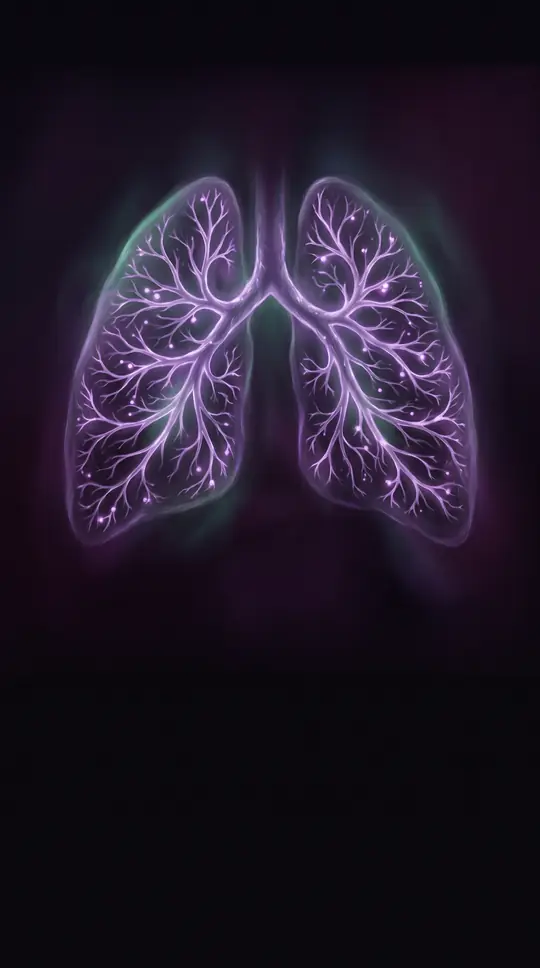
In short: Deep within your lungs, hundreds of millions of microscopic cavities called alveoli perform the essential task of gas exchange. These fragile structures, unique to mammals, provide a massive surface area for oxygen to enter your blood while carbon dioxide is expelled, a process vital for every breath you take.
The human lung is a marvel of biological engineering, containing approximately 480 million alveoli that create a total surface area of up to 80 square metres for gas exchange. These cup-shaped cavities are the functional units of the lung parenchyma. Each alveolus is wrapped in a fine capillary network, separated by a membrane as thin as 0.2 micrometres. This structure is supported by an extracellular matrix of collagen and elastic fibres, which provide the necessary firmness and elasticity for the lungs to expand during inhalation and recoil during exhalation.
Three primary cell types maintain this delicate system. Type I pneumocytes are thin, flat cells that form the structural wall of the alveolus, covering over 95 percent of its surface to facilitate diffusion. Type II pneumocytes are smaller, cuboidal cells that secrete pulmonary surfactant, a fatty substance that reduces surface tension and prevents the alveoli from collapsing. Finally, alveolar macrophages act as mobile immune scavengers, patrolling the internal surfaces to engulf dust, bacteria, and other foreign particles. If the delicate balance of these cells is disrupted—whether by infection, cigarette smoke, or genetic factors—the resulting damage can lead to severe conditions like emphysema, pulmonary edema, or acute respiratory distress syndrome.
The development of these structures is a prolonged process, beginning at 22 days of gestation and continuing until approximately eight years of age. During the first three years, lung growth is driven primarily by the formation of new alveoli, after which both the number and size of these sacs increase. Modern research, including the development of 'alveolus-on-chip' platforms, is now allowing scientists to model these complex epithelial-endothelial interactions. By studying how these cells respond to stressors like cigarette smoke or viral pathogens, researchers hope to better understand and treat the many diseases that compromise this critical interface between air and blood.
Opens the Intellect+ feed with this card first. Swipe up for the next one.
Sources
- Summary by Intellect · source: Pulmonary alveolus
To cite this card, link to https://intellectplus.co.uk/app/health-pulmonary-alveolus.
How we source, check and correct cards: Editorial standards.
General information, not professional advice. Spotted an error? Flag the card or see corrections and complaints.
Intellect Plus is a visual educational discovery platform for exploring fascinating facts and ideas. Brain Food, Not Brain Rot. What is Intellect Plus?
Related
More in Anatomy and morphology · More in Health · Latest · A–Z · All topics
