How animals master the physics of color and camouflage
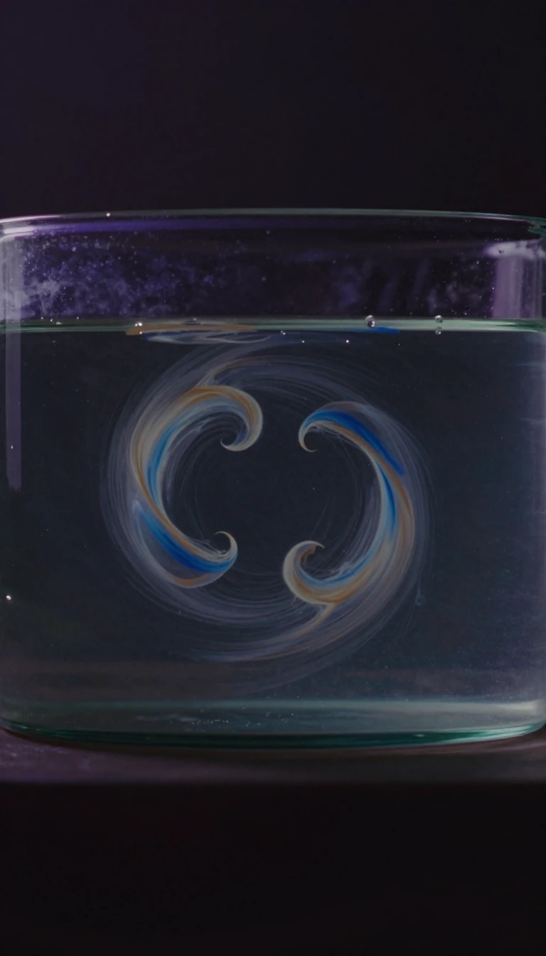
In short: From the shimmering scales of a fish to the sudden shift of an octopus, animals use specialized cells to manipulate light. These aren't just pigments; they are biological machines capable of rearranging structures and moving fluids to create a dynamic, living canvas of color.
Chromatophores are specialized cells found in a wide range of animals, including fish, reptiles, amphibians, and cephalopods. Unlike mammals and birds, which rely primarily on melanocytes, these organisms utilize diverse subclasses of chromatophores to produce color. These include xanthophores (yellow), erythrophores (red), melanophores (black/brown), and leucophores (white). While many of these rely on biochromes—pigments like carotenoids and pteridines that absorb specific wavelengths—others, such as iridophores and leucophores, produce color through structural properties like scattering and optical interference.
The mechanism of color change, known as metachrosis or physiological color change, varies by species. In cephalopods like the octopus, complex organs controlled by muscles allow for rapid shifts. In contrast, vertebrates like chameleons use cell signaling, involving hormones or neurotransmitters, to trigger changes. In many reptiles, these cells form Dermal Chromatophore Units (DCUs), where a basket-like layer of melanophores sits atop iridophores and xanthophores. By aggregating or dispersing melanin, the animal can either hide these underlying layers to appear dark or expose them to create vibrant hues like green, which results from yellow filtering of scattered light.
This biological engineering is so precise that it can be manipulated by external factors. For instance, researchers demonstrated that frogs fed on carotene-restricted crickets lost their red/orange 'filter,' turning them blue. Furthermore, the orientation and thickness of guanine plates in fish iridophores can be regulated by the hormone Acetylcholine. Scientists now study these mechanisms not just to understand animal behavior, but as a sophisticated tool for drug discovery and understanding human disease.
The history of this science dates back to Aristotle's 4th-century BC observations of the octopus. It wasn't until 1819 that Giosuè Sangiovanni first described these cells as 'cromoforo.' Even today, while we can classify these cells by their appearance, the underlying biochemistry—such as the tyrosinase enzyme's role in melanin synthesis—remains the primary focus for understanding how these cells function at a molecular level.
Opens the Intellect+ feed with this card first. Swipe up for the next one.
Sources
- Summary by Intellect · source: Chromatophore
- Also: www.britannica.com — Chromatophore | biological pigment | Britannica
To cite this card, link to https://intellectplus.co.uk/app/science-chromatophore.
How we source, check and correct cards: Editorial standards.
General information, not professional advice. Spotted an error? Flag the card or see corrections and complaints.
Intellect Plus is a visual educational discovery platform for exploring fascinating facts and ideas. Brain Food, Not Brain Rot. What is Intellect Plus?
Related
More in Cells · More in Science · Latest · A–Z · All topics
