Your heart valves are biological one-way gates that keep your blood moving forward
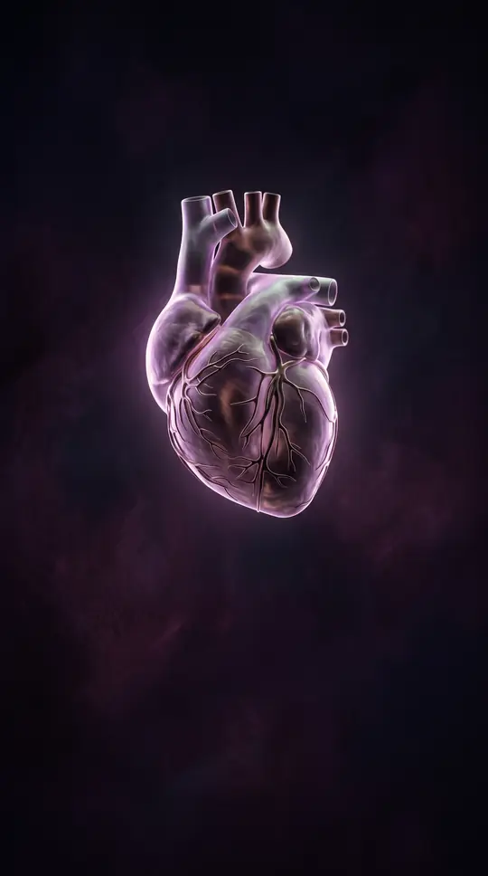
In short: The human heart relies on four precision valves to maintain unidirectional flow. When these structures fail to open fully or close tightly, the heart must work significantly harder to pump blood. Understanding how these gates function—and why they sometimes falter—is essential for recognizing the early signs of disease.
The human heart contains four distinct valves: the aortic and mitral valves on the left side, and the tricuspid and pulmonary valves on the right. These structures function as one-way gates, opening and closing in response to pressure gradients across the heart chambers. The mitral and tricuspid valves, known as atrioventricular valves, separate the upper atria from the lower ventricles. They are anchored by a subvalvular apparatus consisting of chordae tendineae and papillary muscles, which prevent the valves from prolapsing into the atria during contraction. In contrast, the semilunar valves—the aortic and pulmonary—sit at the base of the major arteries, preventing blood from flowing back into the ventricles.
Valve disease occurs when these gates either fail to open completely, a condition called stenosis, or fail to seal properly, known as regurgitation or prolapse. These malfunctions force the heart to exert extra effort to circulate blood. While some individuals are born with congenital defects, others develop valve issues due to aging, high blood pressure, high cholesterol, smoking, or conditions like rheumatic fever and endocarditis. Symptoms can be subtle, including shortness of breath, fatigue, dizziness, or heart palpitations, though many people remain asymptomatic until a doctor detects a heart murmur during a routine examination.
Management depends on the severity of the condition. Mild cases may require only regular monitoring, while severe disease often necessitates medication to manage blood pressure, heart rate, or fluid retention. In advanced cases, surgical intervention is required to repair the damaged valve or replace it with an artificial, human, or animal tissue substitute. Because valve disease can lead to serious complications like heart failure, stroke, or infection, ongoing care is critical. For rare cases, such as carcinoid heart syndrome, surgical replacement is feasible, though long-term durability can be limited by the progression of underlying diseases, often requiring repeat procedures.
Opens the Intellect+ feed with this card first. Swipe up for the next one.
Sources
- Summary by Intellect · source: Heart valve disease
- Also: Acute and midterm outcomes of patients undergoing right-sided heart valve surgery for carcinoid heart valve disease.
- Also: Heart valve
- Also: 4 Heart Valves: What They Are and How They Work
To cite this card, link to https://intellectplus.co.uk/app/health-heart-valve.
How we source, check and correct cards: Editorial standards.
General information, not professional advice. Spotted an error? Flag the card or see corrections and complaints.
Intellect Plus is a visual educational discovery platform for exploring fascinating facts and ideas. Brain Food, Not Brain Rot. What is Intellect Plus?
Related
More in Anatomy and morphology · More in Health · Latest · A–Z · All topics
