Beyond the glass lens: how we see the invisible world of the minute
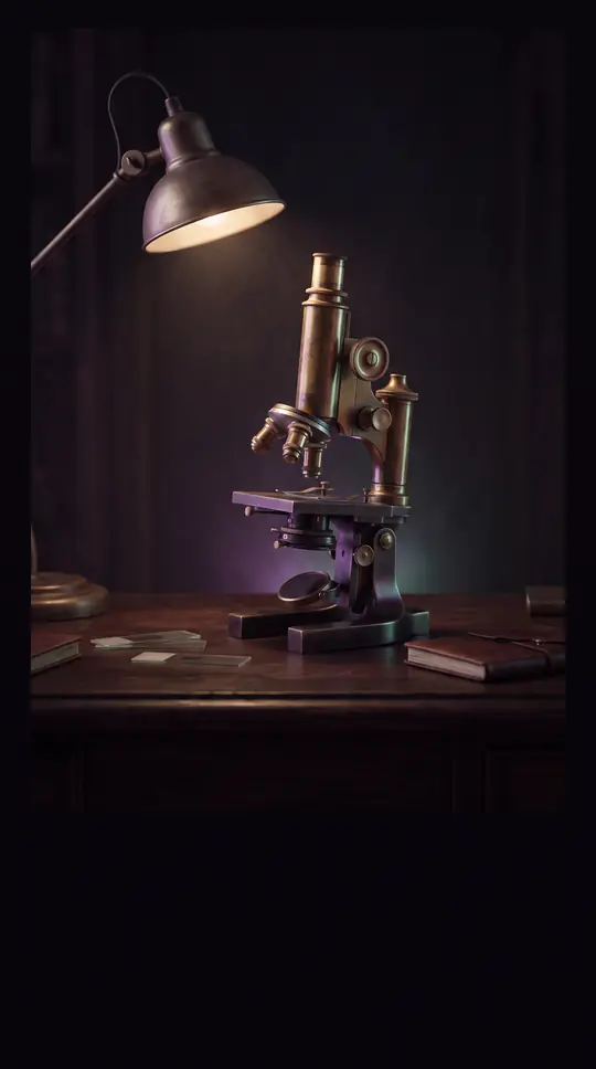
In short: Microscopy is more than just magnifying small objects. From simple handheld lenses to electron beams capable of imaging individual atoms, these instruments allow us to probe structures far beyond human perception. Understanding how they work reveals the sophisticated physics behind our ability to visualize the building blocks of life.
At its core, a microscope is an instrument designed to produce enlarged images of minute structures, bringing them into a scale suitable for human analysis. While we often associate these tools with glass lenses and visible light, the field has evolved to harness various wave forms, including acoustic, X-ray, and electron beams. The power of these devices is measured by two primary metrics: magnification, a dimensionless ratio of how much larger an object appears, and resolution, the smallest detail that can be distinguished.
Optical microscopes remain the most familiar, using glass lenses to manipulate light. A simple microscope, consisting of a single lens, can magnify up to 300 times and resolve details down to one micrometre. Compound microscopes, which use multiple optical components, push this further to 2,000 times magnification and a resolution of about 0.2 micrometres. However, these tools face inherent limitations, particularly when viewing transparent biological samples that lack natural contrast. To overcome this, researchers often use staining techniques, though these can sometimes introduce artifacts that distort the true structure of the specimen.
For even greater detail, scientists turn to electron microscopy. Transmission electron microscopes (TEM) use electron beams in a near vacuum to achieve magnification powers exceeding 1,000,000 times, allowing for the study of thin sections at an atomic level. Meanwhile, scanning electron microscopes (SEM) provide a different perspective by creating reflected images of contoured, solid surfaces. Modern advancements, such as digital imaging and time-lapse micrography, have further democratized these capabilities, moving the field away from traditional eyepieces and toward direct digital observation, making complex imaging tasks accessible to a wider range of practitioners.
Opens the Intellect+ feed with this card first. Swipe up for the next one.
Sources
- Summary by Intellect · source: Microscope | Types, Parts, History, Diagram, & Facts | Britannica
- Also: Microscopy
To cite this card, link to https://intellectplus.co.uk/app/science-microscopy.
How we source, check and correct cards: Editorial standards.
General information, not professional advice. Spotted an error? Flag the card or see corrections and complaints.
Intellect Plus is a visual educational discovery platform for exploring fascinating facts and ideas. Brain Food, Not Brain Rot. What is Intellect Plus?
Related
More in Measurement · More in Science · Latest · A–Z · All topics
