Why the left side of your heart is significantly more muscular than the right
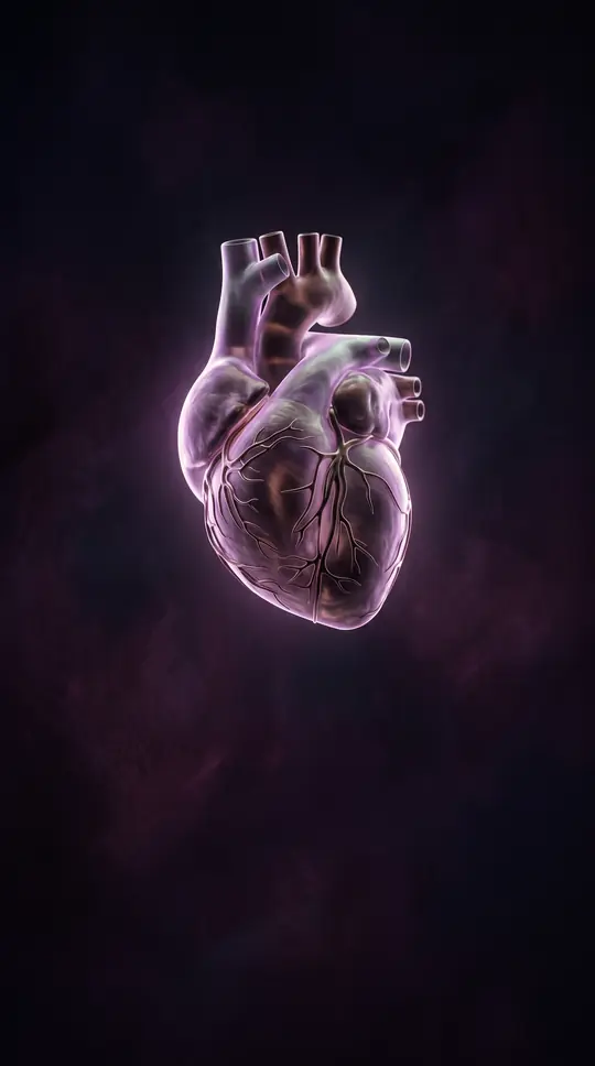
In short: Your heart is a double pump, but its two ventricles are not created equal. While the right ventricle handles the low-pressure task of sending blood to the lungs, the left ventricle must generate massive force to push blood throughout your entire body. This structural disparity is essential for survival.
The human heart functions as a double circulatory system, utilizing two distinct ventricles to manage blood flow. The right ventricle is responsible for pulmonary circulation, directing deoxygenated blood toward the lungs. In contrast, the left ventricle serves the systemic circulation, pumping oxygenated blood through the aorta to the rest of the body. Because the systemic circuit requires significantly higher pressure than the pulmonary circuit, the left ventricle has evolved to be much thicker and more muscular than its right-sided counterpart.
By young adulthood, the walls of the left ventricle are typically three to six times thicker than those of the right. This physical adaptation allows the left ventricle to overcome the high resistance of the systemic arteries. While the right ventricle operates at lower pressures, the left ventricle must push blood into the aorta at a resting pressure of approximately 120 mmHg, compared to the 80 mmHg pressure it receives from the pulmonary veins. This mechanical workload is constant, with the heart pumping roughly 5 liters of blood per minute at rest, a capacity that can surge to 45 liters per minute in elite athletes.
Beyond simple thickness, the ventricles feature complex internal structures. The inner walls are lined with muscular columns known as trabeculae carneae, which include papillary muscles that anchor the heart's valves. The left ventricle is conical and nearly circular in cross-section, whereas the right ventricle appears crescent-shaped and triangular. This anatomical precision is vital; if the conduction system fails, the ventricles can even initiate their own beats, though irregularities like ventricular fibrillation remain the most common cause of sudden cardiac arrest.
The performance of these chambers is monitored through parameters like stroke volume and ejection fraction. Clinicians also track ventricular pressure, as elevated levels can indicate conditions like aortic stenosis. While the heart is remarkably resilient, some individuals are born with only a single functional ventricle, requiring complex surgical interventions like the Fontan procedure to separate circulations and maintain life in the absence of a dedicated sub-pulmonary pump.
Opens the Intellect+ feed with this card first. Swipe up for the next one.
Sources
- Summary by Intellect · source: Ventricle (heart)
- Also: Protocol for non-invasive assessment of skeletal muscle structure and function in adolescents with single ventricle heart disease: a cross-sectional, case-control study.
To cite this card, link to https://intellectplus.co.uk/app/health-ventricle-heart.
How we source, check and correct cards: Editorial standards.
General information, not professional advice. Spotted an error? Flag the card or see corrections and complaints.
Intellect Plus is a visual educational discovery platform for exploring fascinating facts and ideas. Brain Food, Not Brain Rot. What is Intellect Plus?
Related
More in Anatomy and morphology · More in Health · Latest · A–Z · All topics
