Why a 140-year-old staining technique remains a cornerstone of modern medical diagnostics
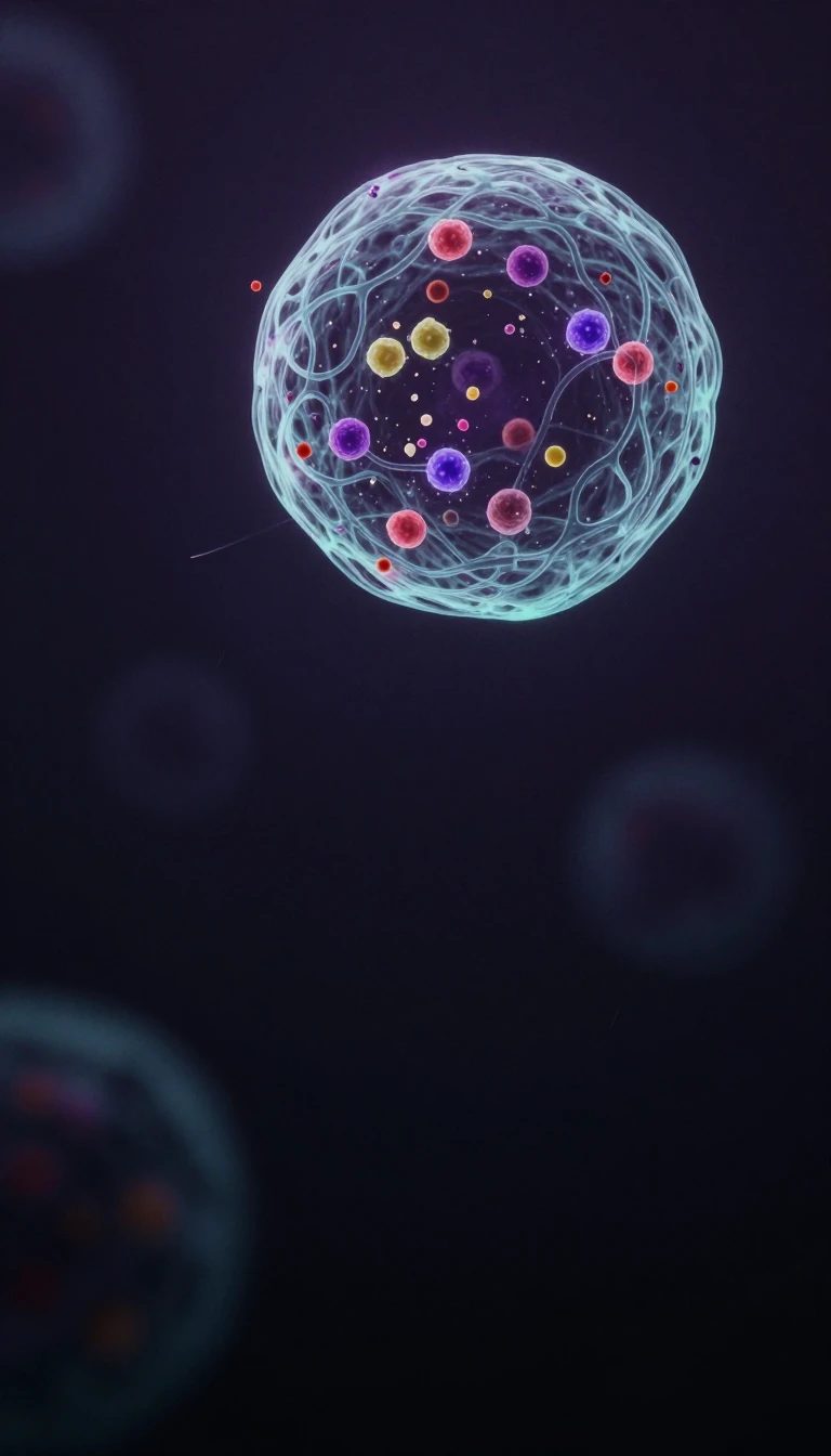
In short: Developed in 1884 to make bacteria visible in lung tissue, the Gram stain remains a primary tool for identifying infections. By exploiting fundamental differences in bacterial cell walls, this simple laboratory test allows scientists to categorize pathogens as gram-positive or gram-negative, providing rapid guidance for life-saving medical treatments.
The Gram stain is a foundational microbiological technique that classifies bacteria based on the chemical and physical properties of their cell walls. Danish bacteriologist Hans Christian Gram introduced the method in 1884 while working in a Berlin hospital morgue. Originally, his goal was not to differentiate bacterial species, but simply to improve the visibility of organisms within stained sections of lung tissue. He observed that certain bacteria resisted decolorization, a discovery that eventually led to the standardized procedure used today.
The mechanism relies on the composition of the bacterial cell envelope. Gram-positive bacteria possess a thick, mesh-like cell wall composed of peptidoglycan, which can make up 50% to 90% of the cell envelope. This structure traps a crystal violet stain, causing the cells to appear purple or blue under a microscope. In contrast, gram-negative bacteria have a much thinner peptidoglycan layer—only about 10% of the wall—and a high lipid content. When treated with a decolorizer like ethanol, the outer membrane of gram-negative bacteria is disrupted, allowing the primary stain to wash away. These cells are then counterstained with safranin, turning them pink or red.
While the test is rapid, it is rarely a standalone diagnostic tool. Healthcare providers typically order it alongside a bacteria culture, which takes several days but provides a definitive identification. The Gram stain serves as a critical triage step, helping clinicians decide on initial treatment paths for conditions like pneumonia, meningitis, or urinary tract infections. Despite its age, the technique remains essential because it provides immediate, actionable data in clinical settings where waiting for a culture could delay necessary care.
Opens the Intellect+ feed with this card first. Swipe up for the next one.
Sources
- Summary by Intellect · source: Gram Stain: What It Is, Purpose, Procedure & Results
- Also: Gram stain | Definition, Procedure, & Facts | Britannica
- Also: Gram stain
To cite this card, link to https://intellectplus.co.uk/app/science-gram-stain.
How we source, check and correct cards: Editorial standards.
General information, not professional advice. Spotted an error? Flag the card or see corrections and complaints.
Intellect Plus is a visual educational discovery platform for exploring fascinating facts and ideas. Brain Food, Not Brain Rot. What is Intellect Plus?
Related
More in Cell biology · More in Science · Latest · A–Z · All topics
